Tandir Orthopedic Implants
Can I have an MRI with orthopedic implants? The answer is often yes, but it is rarely a simple yes-or-no decision. Most modern orthopedic implants use materials that do not move dangerously inside an MRI scanner. However, some implants can heat, distort images, or require specific scanning conditions. The implant’s exact model matters more than the word “orthopedic.”
Frank G. Shellock, PhD, a leading MRI safety expert, has stated, “The presence of an implant does not necessarily mean that a patient cannot undergo an MRI examination.” His point is important. A metal plate, joint replacement, spinal rod, screw, or fixation device should not automatically end the conversation. Instead, the imaging team must check its material, location, age, and safety labeling. MR Conditional devices may be scanned only under defined limits, including magnetic strength, absorption rate, or positioning.
Bring your implant card if you have one. Surgical records, manufacturer details, and previous imaging can also help. Tell the radiology team about every implant, even older hardware that feels harmless. The scanner may produce a loud knocking sound. You may feel nothing unusual, but discomfort should be reported immediately.
The difficult part is incomplete information. A missing implant record can slow the appointment, and that caution is appropriate. Never rely on internet assumptions or a casual verbal approval. A radiologist, MRI technologist, and sometimes your orthopedic specialist should review the case together. This guide examines the ten most important answers, including safety checks, common implant types, image artifacts, warning signs, and questions to ask before entering the scanner.
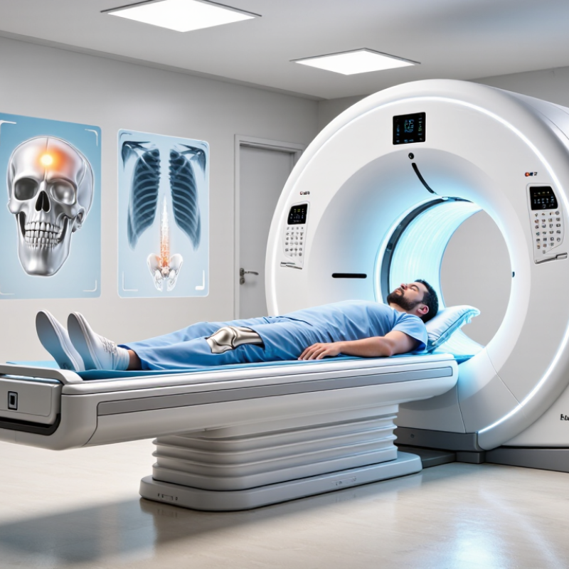
An MRI scan uses a strong magnetic field and radio waves to create detailed images inside the body. It does not use X-rays. The scanner may produce loud knocking sounds, and the examination often lasts between 15 and 60 minutes.
Orthopedic implants can make MRI safety more complicated. Many modern joint replacements, screws, plates, and rods are designed for MRI under specific conditions. However, “metal in the body” is not a complete answer. The implant’s material, location, age, and labeling all matter. Some devices may heat slightly, move, or distort nearby images. The exact risk depends on the scanner and its settings.
Tell the MRI team about every implant, even small screws or surgical clips. Bring implant records if available. The team may ask about the surgery date, body part, and hospital where it occurred. They can check technical documentation before scanning. Do not guess.
The image may still be imperfect.
An implant near the knee, hip, or spine can create bright or dark shadows. These artifacts may hide nearby tissue. A radiologist can adjust the scan or choose another imaging method when necessary. If the implant type is unknown, the appointment may need to pause while records are verified. Never enter the scanner room with loose metal objects, and follow the screening instructions carefully.
Orthopedic implants can affect MRI safety through magnetic attraction, heating, and image artifacts. The exact implant model, material, fixation method, and MRI conditions must be checked before scanning.
Frequencies are calculated using the hydrogen proton gyromagnetic ratio of approximately 42.58 MHz per tesla: 63.87 MHz at 1.5 T and 127.74 MHz at 3 T. These values describe the MRI field, not the safety status of any specific implant.
Metal does not automatically mean an MRI is forbidden. It also does not prove that the scan is safe. Orthopedic implants may be made from titanium, stainless steel, cobalt alloys, or other materials. Their safety depends on the exact device, location, and MRI conditions.
Start with the implant card, surgical report, or hospital records. Look for the precise manufacturer, model, and lot information. Then check its official MRI classification: MR Safe, MR Conditional, or MR Unsafe. “MR Conditional” requires specific limits, such as scanner strength, body position, scan duration, or heating controls. Never rely on a vague note saying “metal implant.”
Tell the radiology team about every implant, including plates, screws, joint replacements, spinal hardware, bone anchors, and older devices. Mention recent surgery, pain near the implant, or missing records. A qualified MRI safety professional can verify the device using clinical databases and manufacturer documentation. X-rays may help identify an unknown implant, but they cannot confirm every safety condition.
The implant may be safe.
Still, details matter. Some implants can move, heat, or create image distortion, even when scanning is allowed. External wires, temporary devices, and abandoned components may require separate assessment. I have seen patients assume that a previous uneventful scan guarantees future safety. That assumption is understandable, but incomplete. MRI approval belongs to the specific implant and scanning protocol, not to the word “metal.”
Most passive orthopedic implants can undergo MRI, but the material alone does not decide safety. Titanium, cobalt-chromium, and many surgical stainless-steel alloys are commonly evaluated for MR environments. Their risks include movement, heating, and image distortion. A 2024 American College of Radiology Manual stresses that implant identification and scanning conditions matter more than appearance.
Look for the exact implant model and its MR labeling. ASTM F2503 recognizes three categories: MR Safe, MR Conditional, and MR Unsafe. MR Conditional devices require specific limits, such as field strength, scan duration, or whole-body SAR. The U.S. Food and Drug Administration also advises checking labeling before exposure. “Metal” is not enough information.
Extra caution is needed with external fixators, magnetic components, programmable systems, and long conductive parts. A titanium joint replacement may be acceptable, while an attached component creates a different risk. Heating can feel like a deep, localized burn. Artifact may also hide tissue around the implant. I would not rely on memory or an old implant card. Bring every available record, even if incomplete. That shortcut is unsafe.
Radiology teams may use the FDA’s MRI safety framework, ASTM testing methods, and ACR screening practices. These sources support a documented, case-specific decision, not a universal yes or no. Some details remain uncertain. Ask the imaging center to verify them before scheduling.
Before an MRI, tell the team about every orthopedic implant in your body. Include joint replacements, plates, screws, rods, pins, and spinal hardware. Share the implant’s name, location, surgery date, and any identification card. Old records can help when your memory is uncertain. Even a confident answer can be incomplete.
Many orthopedic implants are safe under specific MRI conditions. However, safety depends on the material, design, position, and MRI settings. The radiology team may need to verify the implant before scanning. Some hardware can also create shadows that reduce image quality. Tell them if you have pain, warmth, swelling, or unusual sensations near the implant. Do not assume a previous successful scan guarantees every future scan is safe.
Tips: Bring surgical records or implant paperwork. Mention metal fragments from earlier injuries. Tell the team about recent operations, even if the incision looks healed. Answer screening questions carefully. If you are unsure, say so. The technologist and radiologist can check further. During the scan, use the call button if you feel burning, pulling, or discomfort. Safety instructions may feel repetitive, but small details matter.
Top 10 Answers: Can I Have an MRI with Orthopedic Implants?
An orthopedic implant does not automatically prevent an MRI. MRI staff first identify the implant, its material, location, and implantation date. They may review surgical records, implant cards, or previous imaging. Some devices are MRI safe only under specific conditions. These conditions can limit scanner strength, positioning, or scan time. Metal may heat, move, or create image distortion. The team checks each risk carefully.
During the scan, staff use screening questions and safety checks. They explain the emergency call button and ask you to report pain, burning, or unusual pulling. Some patients receive extra padding or careful positioning around the implant. Staff may communicate through an intercom and monitor you throughout the examination. The scan can stop if symptoms appear. The process is careful, but not perfect. Missing implant information can delay safe decisions.
Tips: Bring implant records whenever possible. Tell staff about every surgery, even old ones. Mention new pain, swelling, or skin warmth after the scan. Do not assume a familiar implant is automatically approved. Ask whether the scan uses special limits for your device. Afterward, follow the team’s instructions and contact your healthcare provider if symptoms continue.
| No. | Common Question or Implant | General MRI Status | What MRI Staff Verify Before the Scan | Risk-Management Steps During the Scan | After-Scan Guidance |
|---|---|---|---|---|---|
| 1 | Can most modern orthopedic plates, screws, rods, and joint replacements be scanned? |
Often possible Many passive orthopedic implants are classified as MR Conditional or are considered safe under specified conditions. The exact implant and scanning limits still matter. |
Staff review the implant record, operative report, implant card, or medical records. They confirm the material, location, model, and any manufacturer-specified MRI conditions. | The team selects appropriate magnetic-field strength, scan sequences, and operating limits. The patient is positioned to reduce discomfort and is given an emergency call device. | Most patients can resume normal activities immediately unless their surgical team has provided separate restrictions. Report unusual pain, warmth, swelling, or skin changes. |
| 2 | What about artificial hips, knees, shoulders, or other joint replacements? |
Usually possible Large joint replacements are commonly scanned, although the implant may create image distortion near the joint. |
Staff identify the joint replacement and assess the purpose of the examination. They determine whether metal-related artifact could limit the diagnostic value of the images. | Metal-artifact-reduction techniques may be used when appropriate. Staff monitor for discomfort and stop the scan if significant heating or pain occurs. | No special recovery is normally required. The radiologist explains if artifact affects the interpretation or if additional imaging may be needed. |
| 3 | Are orthopedic screws, pins, wires, or staples a problem? |
Often possible Fixed orthopedic hardware is frequently compatible with MRI, but temporary devices and unidentified metal require individual assessment. |
Staff determine whether the item is internal or external, permanent or temporary, and securely fixed. They pay particular attention to recently placed or unidentified hardware. | The area is monitored for pain, pulling, vibration, or unusual warmth. External metal objects are removed whenever possible. | Patients should follow their surgeon’s wound-care and activity instructions. New or worsening pain near the implant should be reported to a healthcare professional. |
| 4 | Can I have an MRI after fracture fixation or spinal fusion? |
Frequently possible Spinal rods, cages, plates, and screws are often scanned under defined conditions, but the timing of surgery and implant details must be confirmed. |
Staff review surgical history, implant documentation, the scanned body region, and any complications such as infection, wound problems, or suspected hardware loosening. | The team uses positioning and sequences that limit artifact and follows the implant’s approved scanning conditions. Communication is maintained throughout the examination. | MRI does not normally weaken properly fixed orthopedic hardware. Continue the surgeon’s prescribed restrictions and seek advice for severe pain, fever, drainage, or neurological symptoms. |
| 5 | What if the implant was placed recently? |
Requires review A recent operation is not automatically a reason to avoid MRI, but healing status, implant documentation, wound condition, and the clinical need must be evaluated. |
Staff confirm the surgery date, implant type, surgical site, wound condition, and the reason for the MRI. The referring clinician may be consulted when details are incomplete. | Staff avoid unnecessary pressure on the operative area, use careful positioning, and monitor for pain or heat. The scan is stopped if symptoms become concerning. | Follow postoperative instructions rather than assuming the scan changes activity limits. Report increasing redness, drainage, fever, severe swelling, or escalating pain. |
| 6 | Can an MRI be performed with an external fixator or other external orthopedic hardware? |
Case-by-case External frames, clamps, traction equipment, and connecting components may have different MRI restrictions and may not be suitable for every scanner. |
Staff identify every component, confirm its MRI labeling, and coordinate with the orthopedic team. Unverified external equipment is treated as unsafe until assessed. | The team follows the most restrictive component’s conditions and prevents loose or conductive parts from contacting the bore or one another. Continuous communication is essential. | The orthopedic team should inspect the device if there was pain, movement, vibration, or concern about alignment. Do not adjust or remove the frame independently. |
| 7 | Can orthopedic implants heat up during an MRI? |
Small risk under conditions Radiofrequency energy can cause heating, particularly with certain configurations, long conductive components, or scanning conditions outside approved limits. |
Staff check the implant’s conditions, location, dimensions when relevant, and the planned scan settings. They also ask about prior MRI-related heating or discomfort. | The patient must report warmth, burning, pain, or unusual sensations immediately. Staff use padding to prevent skin-to-skin contact and avoid loops or contact with conductive materials. | Heating symptoms usually resolve when scanning stops. Persistent pain, blistering, redness, or a burn-like skin change requires prompt medical evaluation. |
| 8 | Can an implant move or be pulled by the MRI magnet? |
Depends on the implant Securely fixed passive orthopedic implants generally have low displacement risk, but ferromagnetic, loose, temporary, or unidentified objects require special caution. |
Staff establish whether the implant is fixed, loose, or removable and verify its magnetic properties and approved conditions. They also screen for unrelated metal objects. | The patient is kept away from the scanner until screening is complete. Staff use controlled positioning and maintain two-way communication throughout the scan. | New mechanical pain, a sensation of movement, or sudden swelling should be reported immediately, even though serious movement is uncommon with approved fixed implants. |
| 9 | Will orthopedic metal ruin the MRI images? |
Possible image artifact Metal can cause signal loss, distortion, and shading, especially near the implant. This is an image-quality issue rather than proof that the scan is unsafe. |
The radiology team considers the implant’s location and the body area being examined. They decide whether specialized metal-artifact-reduction sequences could improve the images. | Staff optimize patient positioning and select suitable imaging parameters. The scan may take longer when additional sequences are required. | The radiologist reports whether artifact limits interpretation. Alternative imaging or another diagnostic approach may be recommended if important anatomy is obscured. |
| 10 | What should I tell the MRI team before arriving? |
Do not skip screening Never assume an implant is safe based only on its appearance or age. The MRI team must make the final safety assessment. |
Provide the implant name or documentation if available, surgery date and body part, prior imaging complications, pregnancy status when relevant, and any other implanted or retained metal. | Complete the safety questionnaire honestly, remove all removable metal, follow staff instructions, and use the call button if discomfort occurs. Sedation or contrast may require additional precautions. | Most orthopedic-implant MRI examinations require no special aftercare. If contrast or sedation was used, follow the separate instructions provided by the clinical team. |
Many passive orthopedic implants can be scanned under specific conditions. Safety depends on the implant model, location, and MRI settings. Metal alone is not enough information. Not always.
Titanium, cobalt-chromium, and some stainless-steel alloys are often evaluated. They may still cause movement, heating, or image distortion. The exact device matters more than its appearance.
Share the implant model, body location, surgery date, and identification card. Bring surgical records, even if they seem incomplete. My memory may be wrong.
MR Safe means the device presents no known MRI hazard under defined conditions. MR Conditional means scanning is allowed only within stated limits. These limits may include field strength, scan time, or energy exposure.
External fixators, magnetic components, programmable systems, and long conductive parts need careful review. An attached component can change the risk. Do not assume every part behaves identically.
Conductive parts may produce localized heating near the implant. This can feel like deep warmth or a burn. Tell the technologist immediately about pain, warmth, or unusual sensations.
Yes. Metal can create shadows or distortion around the implant. Important tissue may become harder to see. The scan may still be useful, but expectations should remain realistic.
No. Different scanners, settings, or implant combinations can change the risk. Ask the imaging team to verify the device each time. That shortcut is unsafe.
If you are wondering, “Can I have an MRI with orthopedic implants,” the answer depends on the implant’s material, design, location, and safety classification. MRI uses strong magnetic fields and radio waves to create detailed images, so some implants may move, heat up, or interfere with image quality. Many modern orthopedic devices are designed to be MRI-safe or MRI-conditional, but this must be confirmed before the examination.
Before your scan, tell the MRI team about every implant, including joint replacements, plates, screws, rods, pins, bone anchors, and implanted electronic devices. Provide implant records or identification information whenever possible. MRI staff may check the device’s labeling, adjust scan settings, use positioning safeguards, and monitor you during the procedure. Report any unusual warmth, discomfort, pulling, or other sensations immediately. After the scan, staff may also confirm that you feel well and that no follow-up is required. Never assume an implant is safe without professional verification.